What is the Difference Between a Retinal Tear and Retinal Detachment?
Retinal Tear
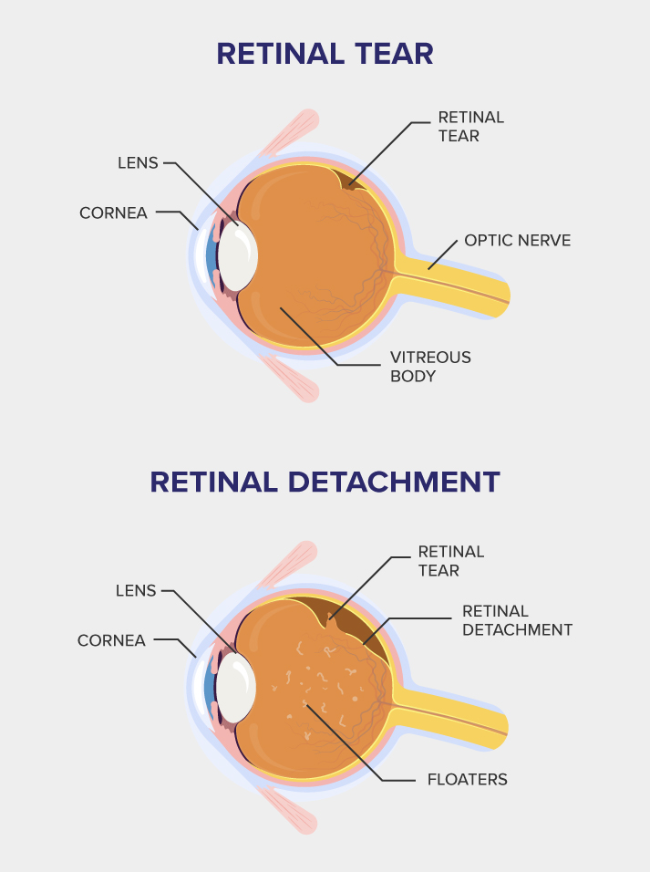
A retinal tear develops when a hole or rip forms in the retina, thelight-sensitive layer of tissue at the back of your eye that captures lightand sends visual signals to the brain.
As we age, the gel-like substance filling the eye (the vitreous) naturallyshrinks and pulls away from the retina. This process is known as aposterior vitreous detachment (PVD) and may occasionally be forcefulenough to tear the retina.
On its own, a retinal tear doesn’t always cause immediate vision loss.However, it can allow fluid to seep beneath the retina, and this mayprogress to retinal detachment if left untreated.
Retinal Detachment
Retinal detachment is a sight-threatening eye emergency that occurswhen the retina separates from the supportive tissue beneath it. Withoutprompt treatment, the retina can lose its blood supply, leading topermanent vision loss.
This condition often begins with a retinal tear, allowing fluid to collectbeneath the retina and causing it to peel away. Those with high myopia,previous eye surgery, eye injuries, or certain retinal conditions face ahigher risk of developing retinal detachment.
Early diagnosis and timely retinal detachment treatment from a specialistgive you the best chance of preserving your sight and preventingblindness.
Symptoms of a Retinal Tear or Retinal Detachment
One of the most important things to understand about a retinal tear or retinal detachment is that both conditions are entirely painless. There’s no discomfort to warnyou that something is wrong, which is exactly why it’s crucial to recognise the visual changes instead.
Symptoms of a Retinal Tear
The symptoms of a retinal tear can come on very suddenly or develop gradually. Since the condition itself is painless, catching the early visual changes is often thebest way to determine that you should seek treatment before more serious effects occur.
You may notice:
- Sudden flashes of light
- New floaters appearing in your vision
- A sudden, rapid increase in floaters
- Blurred vision
- Loss of side (peripheral) vision in some cases
New floaters may appear as:
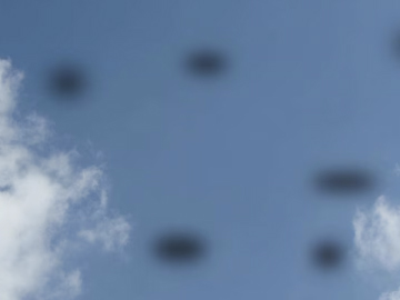
Black spots
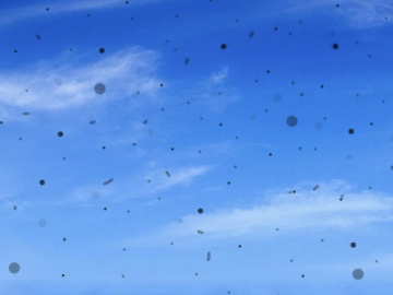
Tiny dots
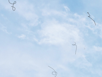
Cobwebs or strings

Tadpole-like shapes
Some people notice only one or two of these signs, while others experience several all at once.
Symptoms of a Retinal Detachment
Unlike a retinal tear, the symptoms of retinal detachment tend to become more pronounced over time, which often makes them easier to notice.
As with a retinal tear, retinal detachment is painless, so it’s the visual changes that you need to watch for:
- Sudden flashes of light
- A sudden increase in floaters
- Noticeably blurry vision
- Loss of side vision
- A grey shadow moving across your field of vision
The “curtain-like” symptom is one of the most important warning signs to act on. If you notice a shadow, curtain, or darkened area moving through your vision, seekemergency care immediately.
Noticing a change in your vision?
If you’ve been experiencing any of these symptoms, it’s important to get a thorough eye examination done. An early diagnosis and timely treatment planfrom an eye specialist can give you the best chance of preserving your vision.
Book An AppointmentWho Is at Risk for Retinal Tear or Detachment?
The most common risk factors for retinal tears and detachment are as follows:
- Myopia (nearsightedness)
- Family history of retinal detachment
- Previous retinal detachment or tears
- Previous eye injury
- Previous eye surgery
- Previous severe eye disease
What Causes Retinal Tear or Detachment?
A retinal tear most often develops when the vitreous gel inside the eye shifts and tugs on the retina over time, eventually causing it to tear. Eye injuries or physicaltrauma can also cause or worsen a retinal tear.
There are three main types of retinal detachment:
1. Rhegmatogenous retinal detachment
The most common type, caused by ageing as the vitreous gel shrinks and tears the retina.
2. Tractional retinal detachment
Caused by scar tissue pulling the retina away from the back of the eye, frequently seen in patients with poorly controlled diabetes (diabetic retinopathy).
3. Exudative retinal detachment
Fluid collects beneath the retina without any accompanying tear, often due to injury, inflammation, or macular degeneration.
Experiencing symptoms of a retinal tear or detachment?
See an eye specialist today
Are you experiencing sudden flashes of light, new floaters, or a dark shadow falling over your field of vision?
These may indicate that you have a retinal tear or detachment, and require urgent evaluation by an eye specialist.
Book an appointment todayWhat Causes Retinal Tear or Detachment?
Diagnosing a retinal tear starts with an eye examination. Your doctor will use eye drops to dilate your pupils, then closely examine the back of your eye using anophthalmoscope and a specialised lens.If a retinal tear or detachment is suspected, your treatment will depend on its size and underlying cause.
Treatment Options for Retinal Tear
Retinal tear treatment often involves laser photocoagulation or cryotherapy. Both procedures create a seal around the tear to help prevent fluid from passingunderneath the retina and reduce the risk of retinal detachment.
Some small retinal tears may not require immediate treatment and can instead be monitored closely through regular follow-up examinations, depending on theirlocation and risk of progressing to retinal detachment.
Treatment Options for Retinal Detachment
Retinal detachment treatment may involve laser photocoagulation or cryotherapy to seal retinal tears, often together with procedures that reposition the retina. Insome cases, a gas bubble may also be injected into the eye to help press the retina back into place while it heals.
There are also three main types of surgeries to repair large retinal tears or detached retinas. Your doctor will choose the most suitable option based on the size of thetear or detachment, and its location in the eye:
1. Pneumatic retinopexy
Your doctor will inject a small gas bubble into the affected eye to push the retina back into place and then use a laser or freezing treatment to seal the tear ordetachment. You will need to hold your head in a specific position for several days to keep the bubble positioned correctly.
2. Scleral buckle
A small, flexible band will be inserted around the white part of the eye to push it inward and toward the retina, helping it reattach. This procedure is performedunder local anaesthesia, and the band remains in place permanently.
3. Vitrectomy
Your doctor will make a small opening in your eye to remove some of the vitreous fluid, and then uses laser, freezing, or a gas bubble to repair the retina.Recovery can take several weeks while the eye heals.
Recovering from Retinal Tear or Detachment
Diagnosing a retinal tear starts with an eye examination. Your doctor will use eye drops to dilate your pupils, then closely examine the back of your eye using anophthalmoscope and a specialised lens.If a retinal tear or detachment is suspected, your treatment will depend on its size and underlying cause.
Treatment Options for Retinal Tear
Retinal tear treatment often involves laser photocoagulation or cryotherapy. Both procedures create a seal around the tear to help prevent fluid from passingunderneath the retina and reduce the risk of retinal detachment.
Some small retinal tears may not require immediate treatment and can instead be monitored closely through regular follow-up examinations, depending on theirlocation and risk of progressing to retinal detachment.
Treatment Options for Retinal Detachment
Retinal detachment treatment may involve laser photocoagulation or cryotherapy to seal retinal tears, often together with procedures that reposition the retina. Insome cases, a gas bubble may also be injected into the eye to help press the retina back into place while it heals.
There are also three main types of surgeries to repair large retinal tears or detached retinas. Your doctor will choose the most suitable option based on the size of thetear or detachment, and its location in the eye:
1. Pneumatic retinopexy
Your doctor will inject a small gas bubble into the affected eye to push the retina back into place and then use a laser or freezing treatment to seal the tear ordetachment. You will need to hold your head in a specific position for several days to keep the bubble positioned correctly.
2. Scleral buckle
A small, flexible band will be inserted around the white part of the eye to push it inward and toward the retina, helping it reattach. This procedure is performedunder local anaesthesia, and the band remains in place permanently.
3. Vitrectomy
Your doctor will make a small opening in your eye to remove some of the vitreous fluid, and then uses laser, freezing, or a gas bubble to repair the retina.Recovery can take several weeks while the eye heals.



