What Is a Macular Hole?
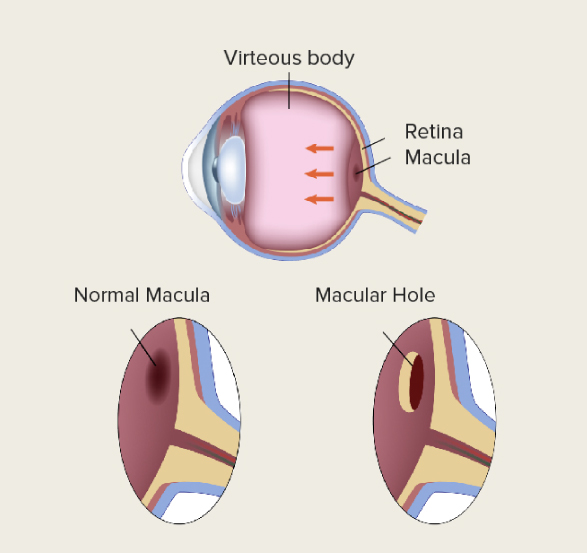
A macular hole is a full-thickness opening that develops in the macula, the central part of the retina at the back of the eye. The macula is responsible for detailed central vision, so a macular hole can affect activities such as reading, recognising faces and seeing fine detail.
BOOK AN APPOINTMENT WITH US TODAY FOR MACULAR HOLE TREATMENTSymptoms of a Macular Hole
Macular holes usually develop in one eye, although they can affect both eyes. They are typically painless, but changes in central vision may become noticeable as the hole develops.
Below are common macular hole symptoms:
- Distorted or wavy vision: Seeing straight lines such as door frames or lines of text looking bent, curved, or wavy.
- Central blurring: Difficulty reading fine print or focusing on detailed objects.
- Reduced visual clarity: Central vision may appear blurry or less detailed in the affected eye.
- Central blind spot: Having a grey, black, or "blank" patch in the central part of your vision.
- Difficulty recognising faces: Difficulty seeing or recognising facial details because central vision is affected.
- Missing or distorted areas: Parts of words or objects may appear distorted, blurred or missing from the centre of your vision.
Noticing Wavy Lines or Blurry Vision?
If you’ve been experiencing any of these symptoms, it’s important to get a thorough eye examination done. An early diagnosis and timely treatment plan from an eye specialist can give you the best chance of preserving your vision.
Book An AppointmentWhat Causes a Macular Hole and Who Is Most at Risk?
Most macular holes are associated with age-related changes in the vitreous, the clear gel that fills the inside of the eye.
With age, the vitreous gradually becomes more liquid and can separate from the retina, a process known as posterior vitreous detachment (PVD). In some eyes, the vitreous remains attached to the macula as it separates from the surrounding retina, creating traction on the macula.
This vitreomacular traction can distort the fovea, the central part of the macula, and in some cases progress to the formation of a macular hole.
Primary Risk Factors for Macular Holes
- Age: Macular holes are most common in older adults, particularly those aged 60 and above.
- Gender: Women are more likely to get the condition than men.
- High cholesterol
Secondary Causes & Contributing Conditions
- High myopia (severe short-sightedness): Highly elongated eyes can be associated with structural changes to the retina and may increase the risk of macular complications, including macular holes.
- Blunt eye trauma: Accidental blows, direct injuries, and similar impact to the eye can cause macular holes.
- Diabetes and diabetic retinal disease: Diabetes and some associated retinal changes have been linked with an increased risk of macular holes.
- Pre-existing retinal conditions: Conditions such as epiretinal membrane, retinal detachment and cystoid macular oedema may be associated with macular hole formation or other macular structural changes.
- Previous eye surgery or inflammation: Previous intraocular surgery and inflammatory eye conditions such as uveitis may be associated with secondary macular holes in some cases.
Experiencing symptoms of a macular hole? See an eye specialist today
Are you experiencing painless central vision blurriness, wavy lines, distorted vision, or a dark spot in the centre of your field of view?
These symptoms may indicate a macular hole and require prompt evaluation by an eye specialist.
Book an appointment todayStages of a Macular Hole
A macular hole can go through several stages of development. The stage is often identified during a professional eye exam.
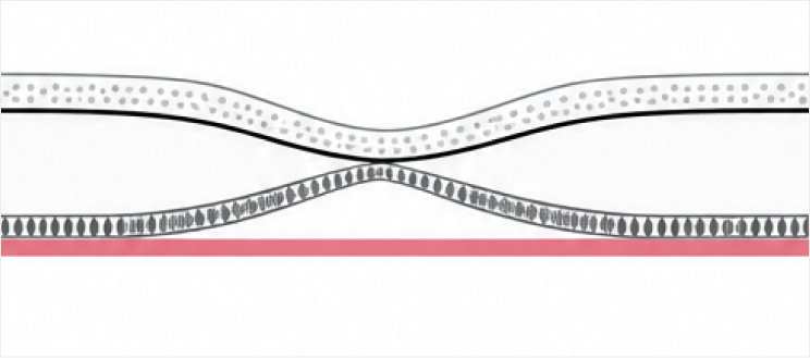
Stage 1: Impending Hole / Foveal Detachment
Stage 1 is an “impending” macular hole in which vitreomacular traction causes changes to the fovea without a full-thickness retinal defect. It may be associated with a small amount of fluid or separation within the foveal tissue.
Symptoms may be mild or absent. Approximately 50% of stage 1 macular holes may close spontaneously, so observation may be appropriate in selected cases.
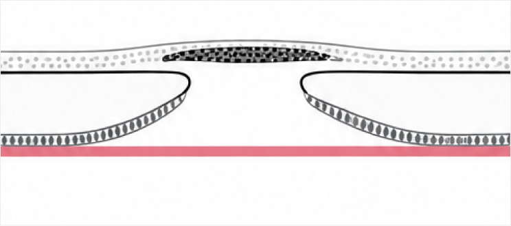
Stage 2: Small Full-Thickness Hole
Stage 2 is a small full-thickness macular hole measuring less than 400 microns in its minimum diameter. The vitreous remains attached to the fovea in most cases.
Central vision may become noticeably blurry or distorted.
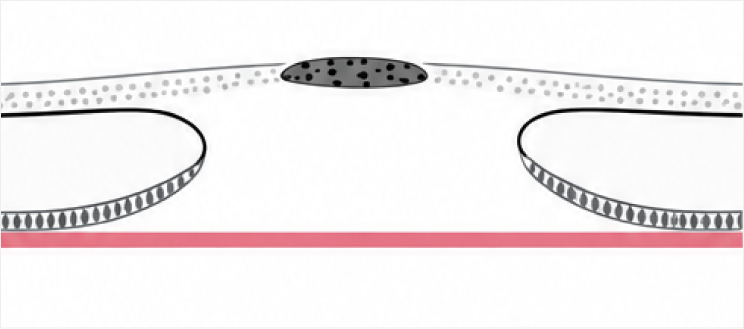
Stage 3: Larger Full-Thickness Hole
Stage 3 is a full-thickness macular hole measuring 400 microns or more. The posterior vitreous is detached over the macula but remains attached elsewhere, and the hole is usually larger than at stage 2.
Central vision loss becomes significant and easy to notice at this stage.
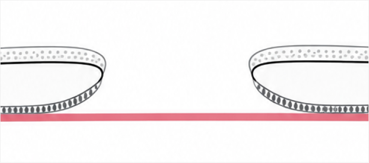
Stage 4: Advanced Hole with Vitreous Separation
Stage 4 is a full-thickness macular hole accompanied by complete posterior vitreous detachment.
The likelihood of successful closure generally depends on factors such as the size and duration of the hole, so your eye specialist can recommend whether observation or surgery is appropriate for your individual case.
How Is a Macular Hole Diagnosed?
Macular holes are diagnosed with a series of tests and examinations carried out by an eye doctor.
- Comprehensive clinical examination
This may involve using a dilated slit-lamp microscope evaluation to examine the retina.
- Optical coherence tomography (OCT)
This non-invasive scan uses reflected light to produce high-resolution cross-sectional images of the retina. It is the main imaging test used to diagnose and stage macular holes and can help assess the size of the hole and associated vitreomacular traction.
- Fundus photography
Photographs of the retina may be taken to document the appearance of the macula and monitor changes over time. Fluorescein angiography is not usually required to diagnose a typical macular hole, but may be performed when your doctor needs to investigate other retinal conditions.
How Is a Macular Hole Treated?
Macular hole treatment depends on the stage, size, duration and characteristics of the hole, as well as its effect on your vision. Options may include observation in selected early cases or vitrectomy surgery for full-thickness macular holes.
How Vitrectomy Surgery Works
- Vitreous removal
Small instruments are inserted through tiny openings in the eye to remove the vitreous gel and release traction on the macula.
- ILM peeling
The surgeon may peel the thin internal limiting membrane (ILM) from the surface of the retina around the macular hole. This helps relieve traction and facilitates closure of the hole.
- Gas bubble insertion
A temporary gas bubble may be placed in the eye to provide gentle pressure over the macula and support closure of the hole while it heals.
Post-Operative Recovery & Head Posturing
- Face-down posturing
If a gas bubble is used, your surgeon may recommend face-down positioning for a period after surgery. The duration varies depending on the size and characteristics of the macular hole and the surgical technique used, and may range from a few days to longer in some cases.
This usually means lying on your stomach or holding your head in a specific position while seated. The goal is to keep the gas bubble in direct contact with your macular tissue for as long as possible.
- Gas absorption
Your vision may remain blurry while the gas bubble is present. The time taken for the gas to dissipate depends on the type of gas used and other factors, and may range from several weeks to longer. Vision can continue to improve after the gas has been absorbed.
- Eye care & medicated drops
You may be prescribed antibiotic and anti-inflammatory eye drops after surgery. Your eye specialist will provide specific instructions on medication use and follow-up care.
An important note
If a gas bubble has been placed in your eye, you must not fly or undergo nitrous oxide anaesthesia until your eye surgeon confirms that the gas has fully dissipated, as changes in pressure can cause a dangerous rise in eye pressure.
Early detection can lead to better treatment outcomes
If you’re experiencing any of the common symptoms for macular holes, our clinic combines diagnostic technology with proven surgical care to customise a treatment plan tailored to your eyes. Take the first step toward preserving your visual clarity today.
Book an appointment todayComplications of Untreated Macular Hole
A macular hole may enlarge or progress over time, potentially causing worsening or permanent changes in central vision. The course varies between individuals, particularly in the early stages.
- Central vision loss
A growing hole may make the centre of your field of vision look blurry, wavy, or blank.
- Central scotoma
A dark or blind spot in your central vision can make tasks like reading or recognising faces harder.
- Permanent distortion
Straight lines can look permanently wavy or bent, a phenomenon called metamorphopsia.
- Full-stage progression
Early or impending macular holes can progress to full-thickness macular holes if vitreomacular traction persists.